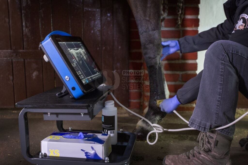
Living with the Draminski BLUE
by Dr Tim Watson BVM&S PhD MRCVS, Waterlane Equine Vets

by Dr Tim Watson BVM&S PhD MRCVS, Waterlane Equine Vets

BLUE is the latest addition to the Polish made range of Draminski ultrasound scanners, offering high-quality images with excelling Doppler ultrasonography for equine and small animal applications. Here Dr Tim Watson, of Waterlane Equine Vets, describes his experiences with the BLUE.
We are a four-vet ambulatory practice that works in Gloucestershire and North Wiltshire. Our client base comprises mainly sports and recreation horses with a substantial amount of polo work in the spring and summer months. Lameness and poor performance investigations occupy two of us on a full-time basis and so access to high-quality imaging is essential.
Earlier this year we started to look at options to replace an ageing Sonosite ultrasound scanner. The remit was for a system that provided outstanding imaging quality, especially when looking at distal limb tendons and ligaments, as well as being easily portable, robust and capable of working off its battery for a full day’s appointments.
Other criteria included quick boot-up – specifically a period that was no longer than either the time taken to prepare the area to be examined or all the small talk I can muster with a nervous owner desperate to know what’s wrong with their horse! It also had to be easy to operate, save and transfer images, and all of this had to be affordable.
We initially looked at second hand GE Logiq and Sonosite Edge scanners but the only systems available within our budget were on eBay and I was nervous about their age and, or, the fact that they came with no warranty. I was nearly seduced by a young and well-appointed GE Logiq until I discovered that, despite being sold as in the UK, it was in fact in Israel and I was to meet the vendor at Heathrow Airport for a demonstration.
It was around this time that our X-ray generator developed a fault. Very obligingly this was late on a Friday just as its supplier was about to close for the weekend and we had several horses requiring urgent radiographs. A client, who is a veterinary nurse, suggested we try Gloucester-based company Photon Surgical Systems and their engineer came to us on a Saturday morning. As I thanked him profusely for getting us back, up and running, he suggested we might look at their latest equine scanner – the Polish made Draminski BLUE.
At a subsequent demonstration, I insisted we test the scanner on what is our hardest ultrasound scenario – hind limb proximal suspensory ligament in large Warmbloods. The BLUE coped with the challenge admirably and even the most sceptical of my colleagues had to agree that the images were outstanding.
We were fortunate to keep the scanner for a week and this also gave me an opportunity to try Draminski’s rectal microconvex probe. We scan a number of mares – for pregnancy diagnosis, artificial insemination or simply because they are difficult – and I was interested to see how this slightly unusual probe compared with more commonplace linear rectal probes.
Nothing in the week-long trial changed our initial impressions of the BLUE. It’s more like a chunky tablet than a traditional ultrasound scanner and would not look out of place with an appropriate fruit logo. It was smaller and lighter than our primary scanner, quicker to boot and proved easy to use, even without referring to the instruction manual. The microconvex rectal probe was also straightforward. So we placed an order and have had the scanner – with 12 MHz linear tendon probe as well as the 6.5 MHz microconvex rectal probe – for just over a month at the time of writing.
So, how have we got on? The BLUE comes in a bespoke hard protective case that is not much larger than a piece of carry-on luggage. The interior is designed so that there are spaces for storage of the mains cable and charger, two probes, stand-off, handily supplied USB stick drive, and fold out stand. The compartments are formed from custom-cut solid foam material, which should prove much more durable than the cut-away customisable foam insert on standard Peli Cases.
The case, fully loaded with scanner and probes et al., weighs in at just 11 kg. One of my colleagues grumbled that there isn’t additional room for surgical spirit, ultrasound gel and wet wipes. But, given that I have previously fatally drowned a scanner in its case with leaking gel and spirit, this is probably a good thing!
The scanner itself weighs just 4 kg and the unit is as robust as it is attractive with an integrated handle. It stands securely by itself and it comes with a detachable foldout stand that makes for a more effective viewing angle. Notably, there are few switches and ports exposed. The on/off button is located on one side with the charging socket and USB, HDMI and Ethernet ports are on the other side behind a sliding cover so they are unlikely to become clogged with straw or shavings.
Probes connect to the rear of the unit and there is an ‘idiot-proof’ label clearly showing which way they should be connected. Boot-up time is a shade over the 25 seconds that the manufacturers quote. Probes can be handily connected and changed without switching the scanner off, and the unit automatically detects which probe is connected.
All functions are touch screen based and include gain, frequency, depth, focus and zoom. There are several pre-sets – of use to us are ‘Horse SDFT’ and ‘Horse DDFT’ – and these actually gave us better images of flexor tendons and suspensory ligaments than our own fiddling with the B mode settings. We have been scanning mares with the ‘Cow Ovary’ pre-set and settings for equine backs, shoulders and feet are included. Pre-sets can be easily adjusted and new ones added. The focus can be moved by simply dragging its marker up or down to the desired depth.
Saving images or cine is straightforward – simply touch the ‘Save Image’ or ‘Save Cine’ button on the screen. This can be done without previously freezing the image – for a rapid save – or after freezing an image that you are happy with. The system then asks for a name (we use the horse’s) and surname (the owner’s). There is a one-touch ability to print or save directly to a USB drive from this screen.
Also on the save screen is an ‘Always ask patient data’ prompt – unclicking this allows subsequent images to be saved without this pop-up screen appearing. But, beware that failing to check this for subsequent images will result in them being stored with the previous patient’s details unless the scanner is switched off between patients, in which case the first image save will ask for patient data.
Saved images are opened via the obvious ‘Open Image’ button and this brings up a screen with each saved image in chronological order with most recent at top, and labelled by name, surname and date/time, along with a thumbnail copy of the image. The latter is particularly helpful in selecting the appropriate image to view or send to USB drive. Multiple images can be clicked and selected for loading or transfer.
A minor grumble with the image archiving software is that each image is saved in a single file, rather than in a patient folder. So, when you touch the ‘Open Image’ button, you may have to scroll through multiple images from numerous patients to find the one you are looking for. Obviously prompt and efficient archiving, following by deleting files, will simplify browsing. Alternatively, individual scans can be located by clicking a ‘filter’ button, which brings up a keypad that enables a search.. (Draminski listened to this and have now updated the software to allow image archiving into patient files.)
There is an extensive set of measuring tools and length can quickly be calculated on a frozen image by touching the screen at the start and end of the tendon, ligament or follicle to be measured without selecting a tool. There are obstetric/gynaecologic tools but these are only for canine and feline reproductive work.
One function that we particularly like is the ability to split the screen horizontally or vertically, or even into four quadrants. Switching to another part of the screen is simply done by clicking that half or quadrant. Another useful feature is being able to enable full-screen viewing of images.
Battery life is good and easily lasts a day’s appointments. The manufacturers quote a life of 2.5 hours continuous use with only a 4-hour charge period. The charging socket doesn’t click or lock in place like some machines we have or tried but the on/off button flashes a blue light to confirm it is connected and charging, even when the unit is switched off.
It’s undoubtedly a little early to comment on the durability of the scanner and probes. The linear tendon probe is solidly constructed with a profile that snuggles into the user’s hand and remains secure even in the presence of ultrasound gel! The cable is over 2 m long, with a protective sleeve and is thick enough so that it doesn’t kink. Frequency is adjustable from 12 MHz down through 10, 9.4, 8 and 7.5 to 6 MHz. Maximum depth is 60 mm.
The tendon probe has performed exceptionally well in evaluating superficial and deep digital flexor tendon lesions, as well as injuries to suspensory, inferior check and annular ligaments. It is very capable of producing excellent images of hind limb proximal suspensory ligaments. I was initially concerned when I scanned a horse with lameness abolished by a deep plantar nerve block and found what looked like a healthy, normal-looking ligament with the BLUE rather than, as I anticipated, a ligament with a disrupted fibre pattern. Further investigation identified substantial osteoarthritis in the centrodistal joint of the hock as the actual source of lameness and this re-affirmed my faith in the scanner.
Colour Doppler is a very welcome bonus in a scanner in this price bracket and is useful in evaluating tendinopathies, especially when monitoring response to treatment and healing. The unit also has Power and Pulse Doppler facilities, although these are arguably more useful in equine cardiac and small animal applications than equine musculoskeletal diseases.
The rectal microconvex probe, which Draminski call the P-probe, has taken a little getting used to. Like the tendon probe, it appears to be of rugged construction, with a thick protective rubber sleeve. The big difference to a convention linear rectal probe is the fact that the microconvex part of the P makes the probe 4 cm in depth, compared with 2-2.4 cm for our other rectal probes. That said it is easy to hold and manoeuvre inside the rectum with grooves on the top and each side for grip. I was a little hesitant at first to use it in small horses and ponies but have found it easy to introduce and well-tolerated.
The microconvex probe seems to make it easier to image ovarian follicles in those horses where ovaries seem to be right at the end of your fingertips when using a linear probe. Adjusting to microconvex as opposed to linear images of the uterus and ovaries has not been a problem. Assessment of uterine oedema, measurement of ovarian follicle size and detection of early pregnancy as well as foetal heartbeat have proved straightforward.
The frequency of this probe is also adjustable, from 9 through 8, 6.5, 5, 4 and 3 to 2 MHz. As well as reproductive work, it has been successfully used to scan chests and abdomens, and for ultrasound-guided injection of sacroiliac joints. I have also used it at home for early pregnancy detection in my suckler cows.
So much for the positives, are there any grumbles? I have already mentioned the image saving function and I am sure it’s a simple software upgrade to allow for archiving in patient files and to enable a search facility. One of the team initially grumbled at the lack of a keyboard but that was quickly overcome when she used the scanner. In fact, the buttons on the touchscreen keyboard are large enough even for my agricultural fingers.
Product support has been good although the real test will come if or when something goes wrong. That said the Draminski BLUE comes with a 2-year warranty and no annual servicing requirements and is reassuringly supported by Photon Surgical System’s in-house team of engineers.
Waterlane Equine Vets, Dr Tim Watson BVM&S PhD MRCVS